
The quest to rewrite human behavior without a scalpel or a pharmaceutical payload has driven neurotechnology into a new, highly aggressive frontier. Transcranial focused ultrasound (tFUS) emerged as the holy grail of non-invasive neuromodulation. By utilizing low-frequency acoustic waves to mechanically gate ion channels deep within the brain, researchers believed they could silently target the nucleus accumbens, quiet the insula, and sever the neurological loops driving substance use disorder (SUD), severe depression, and obsessive-compulsive pathology. It was heralded as a zero-risk, high-precision revolution.
Then, the biological reality check arrived. Reports of localized brain injury, microvascular damage, and adverse neurological events during focused ultrasound neuromodulation trials for SUD shattered the illusion that acoustic energy is inherently benign. The brain is not a passive sponge for sound waves; it is a highly delicate, fluid-filled, heterogeneous matrix encased in an acoustically chaotic skull. When the parameters of low-frequency stimulation cross invisible biophysical thresholds, the mechanical energy intended to modulate a neuron instead tears through the microvasculature.
As a strategist focused on regenerative biology and systems neuroscience, I view this adverse event not as a failure of the technology, but as a profound lesson in biophysics. We must strip away the clinical optimism and look strictly at the acoustic, thermal, and biochemical reality of what happens when sound waves breach the cranial vault. Let’s decode the mechanics of acoustic cavitation, the catastrophic cascade of blood-brain barrier disruption, and the rigorous safety protocols required to salvage the future of non-invasive neuromodulation.
The Physics of Transcranial Focused Ultrasound (tFUS)
To understand how a non-invasive therapy causes physical brain injury, we must first understand how sound becomes a biological signal. Unlike transcranial magnetic stimulation (TMS), which uses magnetic fields to induce electrical currents in the superficial cortex, tFUS utilizes high-frequency sound waves (typically between 200 kHz and 650 kHz for deep brain targets) that can be focused into a millimeter-precise acoustic focal point deep within the parenchyma.
Mechanotransduction and Ion Channel Gating
The intended mechanism of low-intensity focused ultrasound (LIFU) is mechanotransduction. The acoustic radiation force exerts a microscopic physical pressure on the neuronal cell membrane. This mechanical stretch physically pulls open mechanosensitive ion channels—specifically the Piezo1 and Piezo2 channels, as well as TRAAK and TREK-1 potassium channels. According to foundational biophysical models published in Nature Reviews Neuroscience, this transient membrane depolarization alters the firing rate of the targeted neural circuit without generating the massive, synchronous electrical storms seen in TMS or electroconvulsive therapy.
In theory, this allows for the silent, sub-threshold modulation of the mesolimbic dopamine pathway, theoretically reducing the cue-induced cravings that drive substance use disorder.
The Thermal and Mechanical Index Thresholds
The danger arises because acoustic energy does not merely push; it vibrates, and it heats. To prevent tissue damage, the FDA and international acoustic consortia rely on two primary safety metrics:
- The Thermal Index (TI): An estimate of the maximum temperature increase in the tissue. Neuromodulation protocols strictly mandate a TI of less than 1.0, ensuring the tissue temperature does not rise more than 1°C.
- The Mechanical Index (MI): An estimate of the likelihood of non-thermal mechanical damage, specifically acoustic cavitation.
When treating deep brain structures for SUD, researchers must use lower frequencies (e.g., 250 kHz to 500 kHz) because higher frequencies are rapidly absorbed and scattered by the skull. However, lower frequencies possess longer wavelengths and require higher acoustic pressures to achieve the same neuromodulatory effect, pushing the Mechanical Index dangerously close to the threshold of mechanical tissue destruction.
The Adverse Event: Analyzing Brain Injury in SUD Neuromodulation
When clinical trial data or case reports surface detailing “brain injury” during tFUS for addiction, the injury is rarely a macroscopic, catastrophic stroke. Instead, it is a highly localized, insidious microvascular trauma. The biological damage is driven by two primary acoustic phenomena: inertial cavitation and skull-induced standing waves.
Acoustic Cavitation and Microhemorrhages
The most devastating risk of low-frequency ultrasound stimulation is inertial cavitation. The brain parenchyma and the cerebrospinal fluid (CSF) contain microscopic, dissolved gas bodies. When the low-frequency acoustic wave passes through the tissue, the negative pressure phase of the sound wave causes these microscopic gas bubbles to rapidly expand. During the subsequent positive pressure phase, these bubbles violently collapse.
This collapse generates localized shockwaves, micro-jets of fluid traveling at hundreds of meters per second, and extreme, transient spikes in local temperature. According to rigorous acoustic safety data published in Ultrasound in Medicine & Biology, when inertial cavitation occurs near the delicate endothelial lining of the brain’s capillaries, the sheer stress physically ruptures the vessel wall. This results in petechial microhemorrhages—tiny, localized bleeds into the neural tissue.
In the context of an SUD trial targeting the ventral striatum or the insula, a microhemorrhage doesn’t just cause localized tissue death; it triggers a massive, localized neuroinflammatory cascade that can alter mood, exacerbate impulsivity, and induce severe autonomic dysregulation. For a deep dive into how the insula and autonomic networks govern systemic resilience, read our clinical protocol on Autonomic Health and HRV Tracking.
Standing Waves and Skull Attenuation Artifacts
The human skull is an acoustic nightmare. It is not a uniform sphere of bone; it is a heterogeneous, multi-layered composite of cortical and trabecular bone, with highly variable thickness and density. When a planar ultrasound wave enters the skull, the differing speeds of sound through bone versus soft tissue cause severe phase aberration.
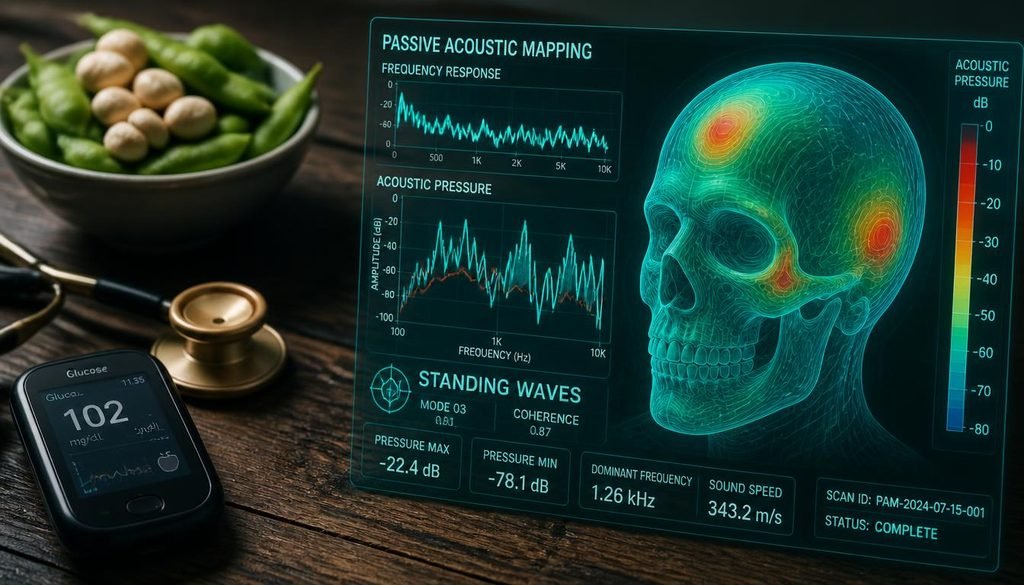
Worse, the inner table of the skull can reflect the acoustic waves back toward the transducer. When the incoming wave and the reflected wave intersect, they create standing waves. In a standing wave, the acoustic pressure at specific nodes (antinodes) can amplify to two or three times the intended intensity. A protocol calibrated to deliver a safe, low-intensity dose to the nucleus accumbens can inadvertently create a localized, high-intensity thermal and mechanical “hotspot” just millimeters away in the overlying cortex or white matter tracts. This localized energy spike is a primary driver of the unintended thermal and mechanical brain injuries observed in complex neuromodulation trials.
Blood-Brain Barrier Disruption: Feature or Fatal Flaw?
One of the most heavily researched applications of focused ultrasound is the intentional, temporary opening of the blood-brain barrier (BBB) to allow chemotherapeutic agents or gene therapies to enter the brain. However, when BBB disruption occurs unintentionally during low-intensity neuromodulation for SUD, it transitions from a therapeutic feature to a fatal biological flaw.
The Biochemical Cascade of Paracellular Leakage
The BBB is maintained by highly specialized endothelial cells locked together by tight junction proteins (claudin-5, occludin, and ZO-1), wrapped in the end-feet of astrocytes. When the acoustic radiation force or micro-cavitation physically stretches these endothelial cells, the tight junctions temporarily unzip, creating paracellular gaps.
If these gaps open in an uncontrolled manner, the results are biochemically catastrophic. The most abundant protein in blood plasma is albumin. When albumin leaks through the compromised BBB into the brain parenchyma, it binds directly to the TGF-β (Transforming Growth Factor-beta) receptors on local astrocytes.
According to landmark neuro-immunological research published in Science Translational Medicine, this albumin-TGF-β binding triggers a massive, maladaptive transcriptional shift in the astrocytes. They cease their supportive, neuroprotective functions and transform into a highly inflammatory, neurotoxic phenotype (the A1 astrocyte). This initiates a vicious cycle of localized excitotoxicity, where the astrocytes fail to clear synaptic glutamate, leading to neuronal overstimulation and subsequent apoptosis.
The Peripheral Immune Invasion
Furthermore, a breached BBB allows peripheral immune cells (macrophages and T-cells) and systemic inflammatory cytokines (IL-6, TNF-alpha) to flood the immune-privileged environment of the brain. This peripheral invasion activates the brain’s resident immune cells, the microglia. Once activated, microglia retract their delicate, synapse-monitoring branches and begin aggressively pruning healthy neural connections while pumping out reactive oxygen species (ROS).
If a patient is undergoing tFUS to cure a substance use disorder, but the acoustic parameters inadvertently breach the BBB and trigger chronic neuroinflammation, the resulting cognitive fog, emotional lability, and depressive crash can severely exacerbate the very addiction pathology the therapy was meant to cure. To understand how systemic, peripheral inflammation breaches neurological barriers, one must look at the parallel mechanisms of intestinal permeability detailed in our guide on Leaky Gut Supplements and Mucosal Repair.
The Microglial Fallout and Oxidative Stress
When microhemorrhages and BBB leakage occur, the secondary injury is often more destructive than the primary acoustic trauma. The extravasation of red blood cells into the neural tissue introduces hemoglobin into the extracellular space.
As the red blood cells lyse, hemoglobin breaks down into heme and free iron. Free iron in the brain is highly toxic. It acts as a potent catalyst for the Fenton reaction, interacting with mild hydrogen peroxide produced by local metabolism to generate the hydroxyl radical—the most destructive reactive oxygen species in human biology.
This localized oxidative stress devastates the lipid bilayers of surrounding neurons (lipid peroxidation) and oxidizes mitochondrial DNA. The damaged mitochondria subsequently fail to produce ATP and begin leaking their own mitochondrial DNA (mtDNA) into the cytoplasm, which is recognized by the cGAS-STING pathway as a viral invader, further amplifying the neuroinflammatory fire.
To halt this secondary neurodegenerative cascade, the brain must rapidly clear the damaged mitochondria and oxidized debris. This requires the upregulation of specific cellular cleanup pathways. For a comprehensive breakdown of how to pharmacologically and behaviorally trigger this cleanup, read our protocol on Next-Gen Mitophagy and Cellular Clearance.
💡 Action Step: The brain’s ability to clear neurotoxic debris, extravasated iron, and misfolded proteins relies entirely on the glymphatic system, which is heavily dependent on optimal cerebrospinal fluid (CSF) hydrodynamics and deep, slow-wave sleep. Chronic dehydration collapses the interstitial space, halting glymphatic flow. Calculate your exact baseline hydration requirements with our Water Intake Calculator to ensure your brain possesses the fluid volume required for nocturnal neuro-immunological clearance.
Safety Protocols and the Future of Non-Invasive Neuromodulation
The occurrence of brain injury during SUD neuromodulation trials is not a death knell for focused ultrasound; it is a mandate for rigorous biophysical oversight. The future of tFUS relies on shifting from “open-loop” acoustic delivery to highly monitored, “closed-loop” systems that can detect tissue damage milliseconds before it becomes permanent.
1. Real-Time MRI Thermometry
The standard of care for high-intensity focused ultrasound (HIFU) ablation is continuous MRI thermometry, which maps the temperature of the brain tissue in real-time. However, low-intensity neuromodulation (LIFU) has historically been performed outside the MRI bore, relying on mathematical models to estimate heating. To eliminate thermal injury, all deep-brain tFUS protocols must be integrated with fast-sequence MRI thermometry, allowing the system to automatically shut off the acoustic power if the local tissue temperature rises by even 0.5°C.
2. Passive Acoustic Mapping (PAM)
To prevent inertial cavitation and microhemorrhages, researchers are deploying Passive Acoustic Mapping. Instead of just emitting sound, the transducer array acts as a highly sensitive microphone, “listening” to the acoustic emissions bouncing back from the brain.
When stable cavitation occurs, the microbubbles emit clean, harmonic frequencies. But the exact millisecond inertial (destructive) cavitation begins, the bubbles emit a broad, chaotic spectrum of broadband noise. By utilizing PAM, the ultrasound system can detect this broadband acoustic signature and terminate the sonication in less than a millisecond, preventing the mechanical shearing of the microvasculature. According to advanced acoustic engineering data published in Physics in Medicine & Biology, PAM is the only reliable method to ensure the Mechanical Index remains strictly below the threshold of vascular damage in heterogeneous human skulls.
3. Patient-Specific Acoustic Ray Tracing
To eliminate standing waves and phase aberration, clinics must abandon “one-size-fits-all” helmet geometries. Prior to a neuromodulation session, the patient must undergo a high-resolution CT scan of the skull. This data is fed into a supercomputer that performs acoustic ray-tracing, calculating the exact phase and amplitude delays required for each individual transducer element to perfectly reconstruct the focal point inside the specific topography of the patient’s skull, effectively canceling out the aberrations and eliminating dangerous cortical hotspots.
4. Systemic Metabolic and Inflammatory Baselines
The brain does not exist in a vacuum; its resilience to acoustic stress is heavily dictated by the systemic biological terrain. A patient with high visceral adiposity, chronic hyperglycemia, and systemic endothelial dysfunction already possesses a fragile, highly permeable blood-brain barrier and a primed, hyper-reactive microglial network. Subjecting this compromised neurovascular unit to acoustic stress drastically lowers the threshold for adverse events.
Before entering a neuromodulation trial, a patient’s systemic inflammatory baseline must be optimized. Track your true metabolic risk and visceral adiposity using our Advanced BMI Calculator and ensure your cellular repair mechanisms are fully fueled by calculating your exact daily amino acid requirements with our Protein Intake Calculator.
Final Thoughts
The translation of focused ultrasound from a tool of physical ablation to a scalpel of behavioral neuromodulation is one of the most exciting frontiers in modern neuroscience. The ability to silently reach into the mesolimbic circuitry and dampen the pathological cravings of substance use disorder holds the potential to save millions of lives.
But biology demands absolute respect for its physical boundaries. The reports of brain injury, microhemorrhage, and blood-brain barrier disruption during low-frequency ultrasound stimulation are stark reminders that acoustic energy is a physical force, not a magic wand. When we ignore the chaotic acoustic reality of the human skull, or push the mechanical index beyond the threshold of microvascular integrity, we trade a behavioral pathology for a neurovascular catastrophe.
By demanding real-time thermometry, passive acoustic mapping, and patient-specific ray tracing, we can tame the acoustic wave. We can harness the profound power of mechanotransduction to rewrite the neural circuits of addiction, while fiercely protecting the delicate, irreplaceable micro-architecture of the human brain.
Frequently Asked Questions (FAQ)
1. How does low-frequency focused ultrasound cause brain injury?
Brain injury during low-frequency ultrasound stimulation is primarily caused by inertial cavitation and standing waves. Inertial cavitation occurs when microscopic gas bubbles in the brain tissue rapidly expand and violently collapse, generating shockwaves that rupture local capillaries, causing microhemorrhages. Standing waves, caused by acoustic reflections off the inner skull, can create localized, high-intensity thermal and mechanical hotspots that damage neural tissue.
2. What happens when focused ultrasound accidentally opens the blood-brain barrier?
Unintentional blood-brain barrier (BBB) disruption allows blood plasma proteins, particularly albumin, to leak into the brain parenchyma. Albumin binds to TGF-beta receptors on astrocytes, triggering them to transform into a highly inflammatory, neurotoxic phenotype (A1 astrocytes). This initiates a cascade of excitotoxicity, neuroinflammation, and localized neuronal damage.
3. Why is the human skull dangerous for ultrasound neuromodulation?
The skull is a heterogeneous, multi-layered bone structure with highly variable thickness and density. This causes severe acoustic phase aberration and scattering. Furthermore, the inner table of the skull can reflect sound waves back, creating “standing waves” that amplify acoustic pressure to dangerous levels in unintended areas of the cortex, leading to localized thermal or mechanical injury.
4. How can doctors prevent brain injury during ultrasound neuromodulation?
Injury prevention requires closed-loop monitoring systems. This includes real-time MRI thermometry to detect microscopic temperature spikes, and Passive Acoustic Mapping (PAM) to “listen” for the broadband acoustic signatures of destructive inertial cavitation, allowing the system to shut off the ultrasound in milliseconds. Additionally, patient-specific CT ray-tracing is used to correct for skull aberrations.
5. Can focused ultrasound cure substance use disorder (SUD)?
Transcranial focused ultrasound is currently an experimental, investigational therapy for SUD. The goal is to use low-intensity acoustic waves to mechanically modulate (quiet) the hyperactive mesolimbic dopamine pathways, such as the nucleus accumbens, thereby reducing cue-induced cravings. While early trials show promise, the risk of microvascular injury and neuroinflammation means it is not yet an approved or standard clinical treatment for addiction.
⚕️ Editorial & Medical Disclaimer
Julian Vance is not a licensed physician or medical professional. All content published on RegenStep.com is thoroughly reviewed against cited primary sources and peer-reviewed literature for educational and informational purposes only. It does not constitute medical advice, diagnosis, or treatment. Always consult a qualified healthcare provider or doctor regarding any personal health condition or medical decisions, particularly before participating in experimental neuromodulation trials, as non-invasive brain stimulation carries significant neurological and vascular risks.

Julian Vance is the writer and editor behind RegenStep, translating peer-reviewed longevity, metabolic, and gut-health research into practical, actionable guides. Julian is not a licensed physician; content is reviewed against cited primary sources and is for informational purposes only. Always consult a qualified healthcare provider for personal medical advice.